

Which Tissue System In Plants Is Most Similar To The Integumentary System In Animals?
| Integumentary system | |
|---|---|
| Cross-section of all pare layers | |
| Details | |
| Identifiers | |
| Latin | inte-meugatary |
| MeSH | D034582 |
| TA98 | A16.0.00.001 |
| TA2 | 7040 |
| TH | H3.12.00.0.00001 |
| FMA | 72979 |
| Anatomical terminology [edit on Wikidata] | |
The integumentary arrangement is the set of organs forming the outermost layer of an beast'southward trunk. It comprises the skin and its appendages, interim equally a concrete barrier betwixt the external environment and the internal environment that it serves to protect and maintain the body of the animal.
The integumentary system includes pilus, scales, feathers, hooves, and nails. It has a variety of additional functions; information technology may serve to maintain water balance, protect the deeper tissues, excrete wastes, and regulate torso temperature, and is the attachment site for sensory receptors to detect pain, sensation, pressure, and temperature.
Structure [edit]
Skin [edit]
The skin is one of the largest organs of the body. In humans, it accounts for about 12 to 15 percent of full trunk weight and covers 1.5-2m2 of surface surface area.[1]

3D still showing human being integumentary system.
The skin (integument) is a composite organ, made up of at least ii major layers of tissue: the epidermis and the dermis.[2] The epidermis is the outermost layer, providing the initial bulwark to the external environs. It is separated from the dermis by the basement membrane (basal lamina and reticular lamina). The epidermis contains melanocytes and gives color to the skin. The deepest layer of the epidermis too contains nerve endings. Below this, the dermis comprises 2 sections, the papillary and reticular layers, and contains connective tissues, vessels, glands, follicles, hair roots, sensory nerve endings, and muscular tissue.[3]
Between the integument and the deep torso musculature in that location is a transitional subcutaneous zone made up of very loose connective and adipose tissue, the hypodermis. Substantial collagen bundles anchor the dermis to the hypodermis in a way that permits almost areas of the skin to move freely over the deeper tissue layers.[4]
Epidermis [edit]
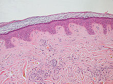
Epidermis and dermis of human skin
The epidermis is the stiff, superficial layer that serves as the first line of protection against the outer environs. The human being epidermis is equanimous of stratified squamous epithelial cells, which further intermission downwards into four to v layers: the stratum corneum, stratum granulosum, stratum spinosum and stratum basale. Where the skin is thicker, such every bit in the palms and soles, there is an extra layer of skin betwixt the stratum corneum and the stratum granulosum, called the stratum lucidum. The epidermis is regenerated from the stalk cells found in the basal layer that develop into the corneum. The epidermis itself is devoid of claret supply and draws its nutrition from its underlying dermis.[5]
Its main functions are protection, assimilation of nutrients, and homeostasis. In structure, information technology consists of a keratinized stratified squamous epithelium; four types of cells: keratinocytes, melanocytes, Merkel cells, and Langerhans cells.
The predominant jail cell type of the epidermis is the keratinocyte, which produces keratin, a fibrous poly peptide that aids in skin protection, and is responsible for the formation of the epidermal water barrier by making and secreting lipids.[6] The majority of the skin on the human being body is keratinized, with the exception of the lining of mucous membranes, such equally the within of the mouth. Non-keratinized cells allow h2o to "stay" atop the construction.
The poly peptide keratin stiffens epidermal tissue to class fingernails. Nails abound from a thin area chosen the nail matrix at an average of 1 mm per week. The lunula is the crescent-shape expanse at the base of operations of the boom, lighter in color as it mixes with matrix cells. Merely primates have nails. In other vertebrates, the keratinizing system at the terminus of each digit produces claws or hooves.[2]
The epidermis of vertebrates is surrounded by two kinds of coverings, which are produced by the epidermis itself. In fish and aquatic amphibians, it is a thin mucus layer that is constantly being replaced. In terrestrial vertebrates, it is the stratum corneum (dead keratinized cells). The epidermis is, to some degree, glandular in all vertebrates, but more than then in fish and amphibians. Multicellular epidermal glands penetrate the dermis, where they are surrounded by blood capillaries that provide nutrients and, in the case of endocrine glands, transport their products.[vii]
Dermis [edit]
The dermis is the underlying connective tissue layer that supports the epidermis. Information technology is composed of dumbo irregular connective tissue and areolar connective tissue such as a collagen with elastin bundled in a diffusely bundled and woven pattern.
The dermis has two layers: the papillary dermis and the reticular layer. The papillary layer is the superficial layer that forms finger-like projections into the epidermis (dermal papillae),[v] and consists of highly vascularized, loose connective tissue. The reticular layer is the deep layer of the dermis and consists of the dense irregular connective tissue. These layers serve to requite elasticity to the integument, allowing stretching and conferring flexibility, while also resisting distortions, wrinkling, and sagging.[3] The dermal layer provides a site for the endings of claret vessels and fretfulness. Many chromatophores are likewise stored in this layer, as are the bases of integumental structures such every bit hair, feathers, and glands.
Hypodermis [edit]
The hypodermis, otherwise known every bit the subcutaneous layer, is a layer beneath the pare. Information technology invaginates into the dermis and is attached to the latter, immediately above it, by collagen and elastin fibers. It is essentially composed of a type of cell known equally adipocytes, which are specialized in accumulating and storing fats. These cells are grouped together in lobules separated past connective tissue.
The hypodermis acts equally an free energy reserve. The fats independent in the adipocytes can be put back into circulation, via the venous route, during intense try or when in that location is a lack of energy-providing substances, and are and then transformed into free energy. The hypodermis participates, passively at least, in thermoregulation since fat is a oestrus insulator.
Functions [edit]
The integumentary system has multiple roles in maintaining the body's equilibrium. All body systems work in an interconnected fashion to maintain the internal conditions essential to the part of the body. The pare has an important chore of protecting the body and acts as the body'southward commencement line of defense against infection, temperature change, and other challenges to homeostasis.[8] [ix]
Its principal functions include:
- Protect the body's internal living tissues and organs
- Protect against invasion past infectious organisms
- Protect the body from dehydration
- Protect the body against abrupt changes in temperature, maintain homeostasis
- Help excrete waste product materials through perspiration
- Act every bit a receptor for affect, pressure, pain, heat, and common cold (run into Somatosensory system)
- Protect the torso against sunburns by secreting melanin
- Generate vitamin D through exposure to ultraviolet light
- Store water, fat, glucose, vitamin D
- Maintenance of the body form
- Formation of new cells from stratum germinativum to repair pocket-size injuries
- Protect from UV rays.
- Regulates body temperature
- Information technology distinguishes, separates, and protects the organism from its surroundings.
Small-bodied invertebrates of aquatic or continually moist habitats respire using the outer layer (integument). This gas exchange system, where gases but diffuse into and out of the interstitial fluid, is called integumentary exchange.
Clinical significance [edit]
Possible diseases and injuries to the human integumentary system include:
- Rash
- Yeast
- Athlete's foot
- Infection
- Sunburn
- Pare cancer
- Albinism
- Acne
- Herpes
- Herpes labialis, commonly called cold sores
- Impetigo
- Rubella
- Cancer
- Psoriasis
- Rabies
- Rosacea
- Atopic dermatitis
- Eczema
References [edit]
- ^ Martini, Frederic; Nath, Judi L. (2009). Fundamentals of anatomy & physiology (eighth ed.). San Francisco: Pearson/Benjamin Cummings. p. 158. ISBN978-0321505897.
- ^ a b Kardong, Kenneth V. (2019). Vertebrates : comparative anatomy, function, evolution (Eighth ed.). New York, NY. pp. 212–214. ISBN978-1-259-70091-0.
- ^ a b "The Ageing Skin – Function 1 – Construction of Skin". pharmaxchange.info.
- ^ Pratt, Rebecca. "Integument". AnatomyOne. Amirsys, Inc. Retrieved 2012-09-28 .
- ^ a b Kim, Joyce Y.; Dao, Harry. "Physiology, Integument". StatPearls. StatPearls Publishing.
- ^ Yousef, Hani; Alhajj, Mandy; Sharma, Sandeep. "Anatomy, Skin (Integument), Epidermis". StatPearls. StatPearls Publishing.
- ^ Quay, Wilbur B. (1 February 1972). "Integument and the Environment Glandular Composition, Function, and Development". Integrative and Comparative Biology. 12 (i): 95–108.
- ^ Integumentary+System at the US National Library of Medicine Medical Subject area Headings (MeSH)
- ^ Marieb, Elaine; Hoehn, Katja (2007). Human Anatomy & Physiology (7th ed.). Pearson Benjamin Cummings. p. 142.
External links [edit]
Source: https://en.wikipedia.org/wiki/Integumentary_system
Posted by: campbellcrusuppeas.blogspot.com
0 Response to "Which Tissue System In Plants Is Most Similar To The Integumentary System In Animals?"
Post a Comment